Articles
- Page Path
- HOME > Res Vestib Sci > Volume 16(2); 2017 > Article
-
Case Report
수평전정안구반사 소실이 주증상인 베르니케뇌병증 4예 - 장혁수1, 신병수1,2, 서만욱1,2, 오선영1,2
- Four Cases of Wernicke’s Encephalopathy with Impaired Horizontal Vestibular Ocular Reflexes
- Hyuk-Su Jang1, Byoung-Soo Shin1,2, Man-Wook Seo1,2, Sun-Young Oh1,2
-
Research in Vestibular Science 2017;16(2):57-63.
DOI: https://doi.org/10.21790/rvs.2017.16.2.57
Published online: June 15, 2017
1Department of Neurology, Chonbuk National University Hospital, Jeonju, Korea
2Department of Neurology, Chonbuk National University Medical School, Jeonju, Korea
- Corresponding Author: Sun-Young Oh Department of Neurology, Chonbuk National University Hospital, Chonbuk National University Medical School, 20 Geonji-ro, Deokjin-gu, Jeonju 54907, Korea Tel: +82-63-250-1896 Fax: +82-63-251-9363 E-mail: ohsun@jbnu.ac.kr
• Received: April 24, 2017 • Revised: May 15, 2017 • Accepted: June 2, 2017
Copyright © 2017 by The Korean Balance Society. All rights reserved.
This is an open access article distributed under the terms of the Creative Commons Attribution Non-Commercial License (http://creativecommons.org/licenses/by-nc/4.0) which permits unrestricted non-commercial use, distribution, and reproduction in any medium, provided the original work is properly cited.
- 8,100 Views
- 218 Download
Abstract
- Wernicke’s encephalopathy (WE) is a neurological disorder induced by a dietary vitamin B1 (thiamine) deficiency which is characterized by encephalopathy, gait ataxia, and variant ocular motor dysfunction. In addition to these classical signs of WE, a loss of the horizontal vestibulo-ocular reflex (VOR) is being reported as the major underdiagnosed symptoms in WE. In this retrospective single center study, we report four cases of WE initially presented with impaired horizontal VOR in addition to the classical clinical presentations, and imaging and neurotological laboratory findings were described.
서 론
증 례
고 찰
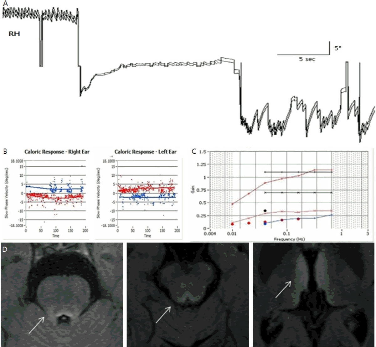
Fig. 1.The patient’s findings (Case 1) of the vestibular function test (A, B, C) and the brain magnetic resonance imaging (MRI) (D). (A) Three-dimensional video-oculography (SMI, Netherland) showed gaze-evoked nystagmus (GEN) during bilateral horizontal gazes. (B) Bithermal caloric tests in the patient showed minimal responses in both ears. (C) Sinusoidal rotation chair test revealed low gain (Gain mean 0.128). (D) Flair (fluid attenuated inversion recovery) MRIs of patient showed hyperintense lesions at both medial thalmi and periaqueductal gray matter. RH, horizontal movement of the right eye.

Fig. 2.The patient’s findings (Case 2) of the vestibular function test (A, B) and the brain magnetic resonance imaging (MRI) (C). (A) Bithermal caloric tests in the patient showed no responses during bithermal caloric irrigation of either ear. (B) Sinusoidal rotation chair test revealed low gain (gain mean 0.073) and increased phase lead of VOR. (C) Flair (fluid attenuated inversion recovery) MRIs of patient showed hyperintense lesions at both mamillary body. VOR, vestibulo-ocular reflex.
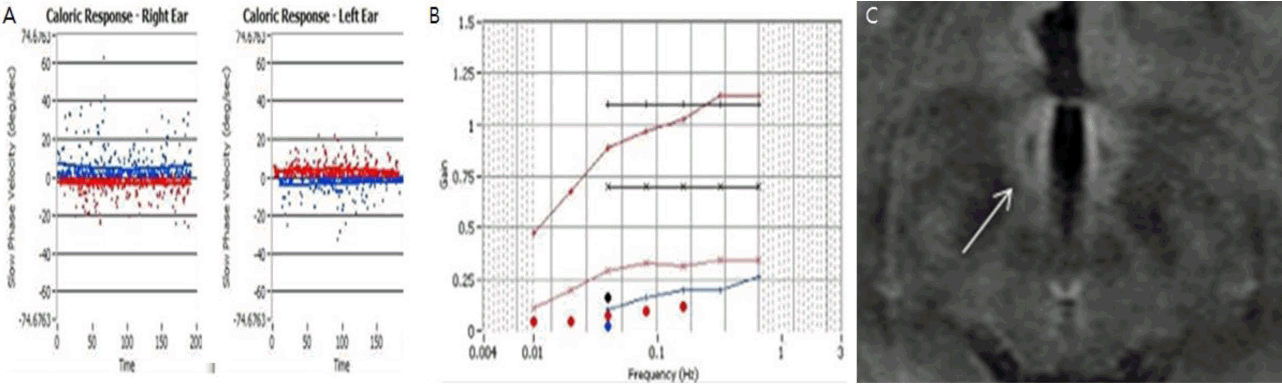
Fig. 3.The patient’s findings (Case 3) of the vestibular function test (A, B, C) and the brain magnetic resonance imaging (MRI) (D). (A) Three-dimensional video-oculography (SMI, Netherland) showed gaze-evoked nystagmus (GEN) during bilateral horizontal gazes. (B) Bithermal caloric tests in the patient showed minimal responses in both ears. (C) Sinusoidal rotation chair test revealed low gain (gain mean 0.195). (D) FLAIR (fluid attenuated inversion recovery) MRIs of patient revealed symmetrical hyperintense lesions at dorsal portions of the medulla, pons, and periaqueductal gray matter. RH, horizontal movement of the right eye.

Fig. 4.The patient’s findings (Case 4) of video head impulse test and brain magnetic resonance imaging (MRI). (A) Video head impulse test showed bilateral catch up saccade on both side. (B) Flair (fluid attenuated inversion recovery) MRIs of patient showed mild hyperintense lesions at both mamillary body and thalamus.
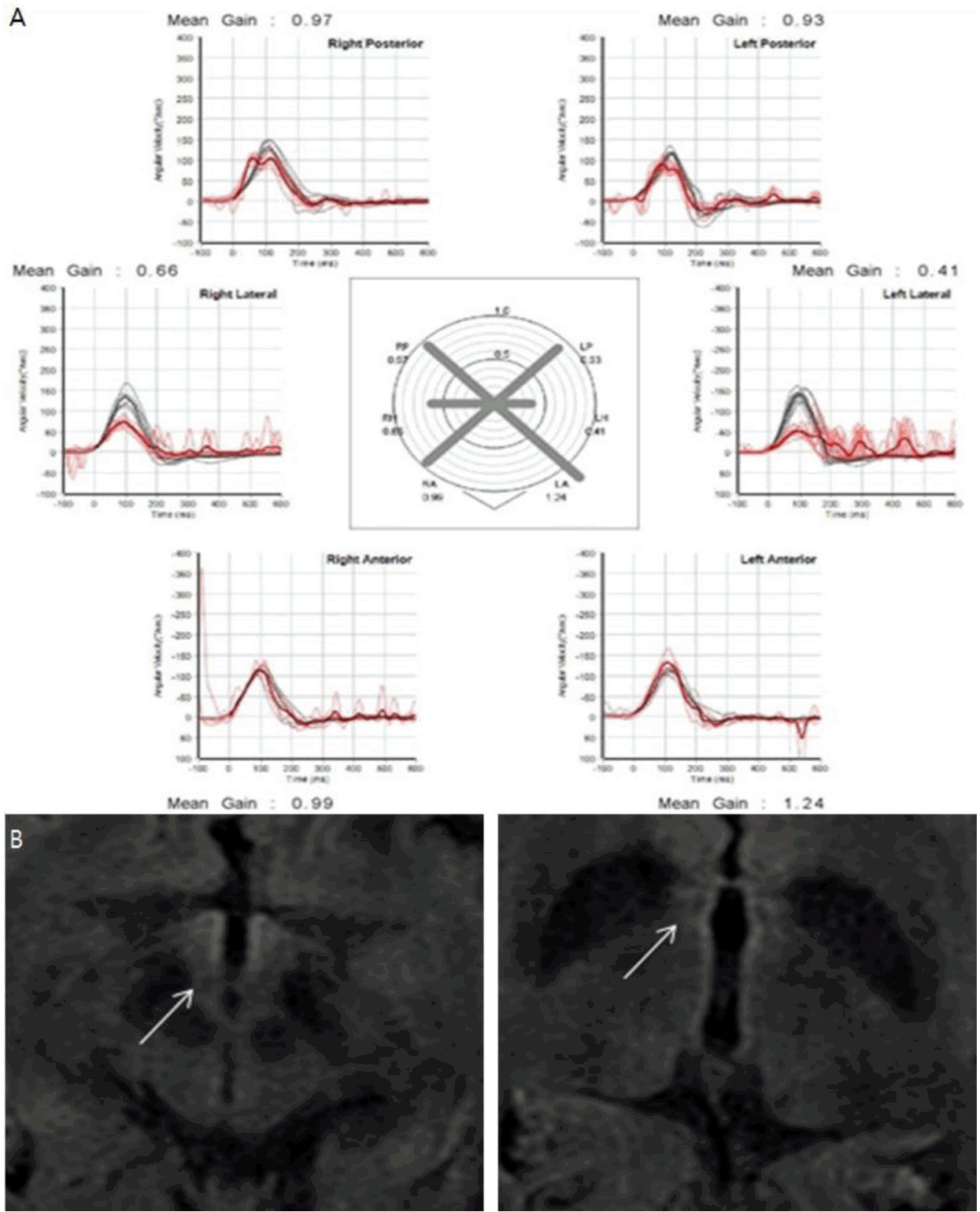
Table 1.Clinical findings of four cases with Wernicke’s encephalopathy
- 1. De La Paz MA, Chung SM, McCrary JA 3rd. Bilateral internuclear ophthalmoplegia in a patient with Wernicke’s encephalopathy. J Clin Neuroophthalmol 1992;12:116–20.PubMed
- 2. Halavaara J, Brander A, Lyytinen J, Setala K, Kallela M. Wernicke’s encephalopathy: is diffusion-weighted MRI useful? Neuroradiology 2003;45:519–23.ArticlePubMed
- 3. Shin BS, Oh SY, Kim JS, Lee H, Kim EJ, Hwang SB. Upbeat nystagmus changes to downbeat nystagmus with upward gaze in a patient with Wernicke’s encephalopathy. J Neurol Sci 2010;298:145–7.ArticlePubMed
- 4. Streifler J, Manor R, Kuritzky A. Ocular manifestations in a case of Wernicke’s encephalopathy caused by hyperemesis gravidarum. Neuro-Ophthalmology 1989;9:267–9.Article
- 5. Sechi G, Serra A. Wernicke’s encephalopathy: new clinical settings and recent advances in diagnosis and management. Lancet Neurol 2007;6:442–55.ArticlePubMed
- 6. Zuccoli G, Pipitone N. Neuroimaging findings in acute Wernicke’s encephalopathy: review of the literature. AJR Am J Roentgenol 2009;192:501–8.ArticlePubMed
- 7. Yang TH, Oh SY. Loss of the horizontal vestibulo-ocular reflex in a patient with wernicke encephalopathy. J Korean Neurol Assoc 2014;32:50–2.
- 8. Cook CC. Prevention and treatment of Wernicke-Korsakoff syndrome. Alcohol Alcohol Suppl 2000;35:19–20.ArticlePubMed
- 9. Ghez C. Vestibular paresis: a clinical feature of Wernicke’s disease. J Neurol Neurosurg Psychiatry 1969;32:134–9.ArticlePubMedPMC
- 10. Choi KD, Oh SY, Kim HJ, Kim JS. The vestibulo-ocular reflexes during head impulse in Wernicke’s encephalopathy. J Neurol Neurosurg Psychiatry 2007;78:1161–2.ArticlePubMedPMC
REFERENCES
Figure & Data
References
Citations
Citations to this article as recorded by 
 PubReader
 ePub Link
 Cite
Cite