소뇌교뇌각 신경초종을 동반한 Brun 안진
Brun’s Nystagmus with Cerebellopontine Angle Schwannoma
Article information

A 70-year-old woman with trigeminal cystic schwannoma developed imbalance and dizziness over a year and a half. Neurologic examination revealed mild abduction limitation of the right eye and positive head impulse test toward the rightward and right peripheral facial palsy.
Videooculography showed a large-amplitude right-beating nystagmus with the rightward gaze and a small-amplitude left-beating nystagmus with the leftward gaze (Supplementary Video 1). Brain magnetic resonance imaging with enhancement demonstrated a well-defined huge mass (about 7×4 cm) in the right cerebellopontine angle compressing the middle cerebellar peduncle and pons (Fig. 1).
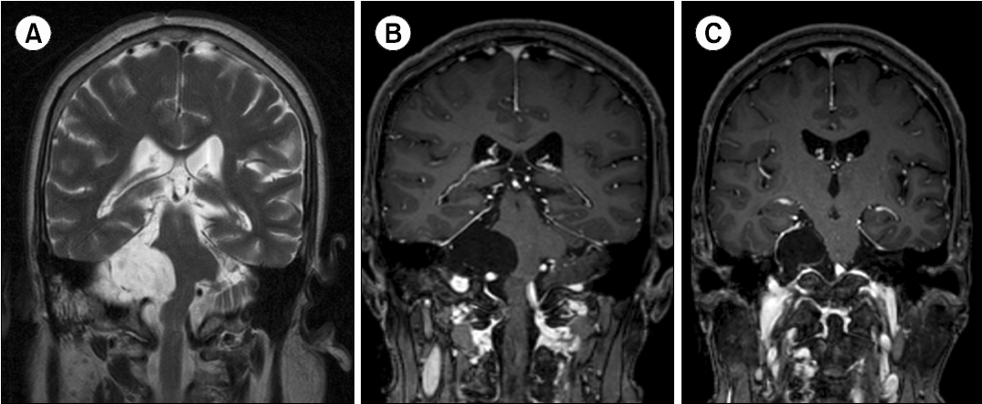
Brain magnetic resonance imaging with enhancement demonstrated a well-defined huge mass (about 7×4 cm) in the right cerebellopontine angle compressing the middle cerebellar peduncle and pons. The mass is high signal intensity on the T2-weighted image (A) and low signal intensity with mild ring enhancement on the T1-weighted images (B, C).
Brun’s nystagmus is a combination of ipsilesional gaze-evoked nystagmus with decreasing velocity waveform and contralesional gaze-induced vestibular nystagmus with linear velocity waveform [1]. The ipsilesional large-amplitude slow nystagmus is the result of impairment of gaze holding due to compression or ischemia of the horizontal neural integrator [2]. The contralesional small-amplitude fast nystagmus is caused by the impairment of the ipsilateral vestibular system. Since this rare directional changing nystagmus indicates localization and lateralization of a lesion, we must know the clinical meaning of this.
Notes
CONFLICT OF INTEREST
Ji-Yun Park is the Editor-in-Chief of Research in Vestibular Science and was not involved in the review process of this article. All authors have no other conflicts of interest to declare.
FUNDING/SUPPORT
None.
AUTHOR CONTRIBUTIONS
Conceptualization: SGH, JYP; Data curation: SGH; Formal analysis: SGH, JYP; Investigation: SGH; Methodology: JYP; Project administration: SJC, MJK; Supervision : SGH, JYP; Visualization: SJC, MJK; Writing–Original Draft: SGH, JYP; Writing–Review & Editing: All authors.
All authors read and approved the final manuscript.
SUPPLEMENTARY MATERIALS
Supplementary Video 1 can be found via https://doi.org/10.21790/rvs.2023.22.2.57. Videooculography showed a largeamplitude right-beating nystagmus with the rightward gaze and a small-amplitude left-beating nystagmus with the leftward gaze. Written informed consent was obtained for publication of this report and accompanying video.