Articles
- Page Path
- HOME > Res Vestib Sci > Volume 18(1); 2019 > Article
-
Case Report
우측전정신경염 환자에서의 시공간기능부전 -
전승호1, 김고운1, 신현준1,2, 신병수1,2, 서만욱1,2, 오선영1,2

- Visuospatial Dysfunction in Patients With the Right Vestibular Neuritis
-
Seung-Ho Jeon1, Ko-Woon Kim1, Hyun-June Shin1,2, Byoung-Soo Shin1,2, Man-Wook Seo1,2, Sun-Young Oh1,2

-
Research in Vestibular Science 2019;18(1):19-23.
DOI: https://doi.org/10.21790/rvs.2019.18.1.19
Published online: March 15, 2019
1Department of Neurology, Chonbuk National University Hospital, Chonbuk National University Medical School, Jeonju, Korea
2Biomedical Research Institute, Chonbuk National University, Jeonju, Korea
- Corresponding Author: Sun-Young Oh Department of Neurology, Chonbuk National University Hospital, 20 Geonji-ro, Deokjin-gu, Jeonju 54907, Korea Tel: +82-63-250-1896 Fax: +82-63-251-9363 E-mail: ohsun@jbnu.ac.kr
• Received: August 3, 2018 • Revised: September 22, 2018 • Accepted: October 1, 2018
Copyright © 2019 by The Korean Balance Society. All rights reserved.
This is an open access article distributed under the terms of the Creative Commons Attribution Non-Commercial License (http://creativecommons.org/licenses/by-nc/4.0) which permits unrestricted non-commercial use, distribution, and reproduction in any medium, provided the original work is properly cited.
- 5,406 Views
- 85 Download
- 1 Crossref
Abstract
- Acute vestibular neuritis (VN) is characterized by acute/subacute vertigo with spontaneous nystagmus and unilateral loss of semicircular canal function. Vestibular system in human is represented in the brain bilaterally with functional asymmetries of the right hemispheric dominance in the right handers. Spatial working memory entails the ability to keep spatial information active in working memory over a short period of time which is also known as the right hemispheric dominance. Three patients (patient 1, 32-year-old female; patient 2, 18-year-old male; patient 3, 63-year-old male) suffered from acute onset of severe vertigo, nausea and vomiting. Patients 1 and 2’s examination revealed VN on the right side showing spontaneous left beating nystagmus and impaired vestibular ocular reflex on the right side in video head-impulse and caloric tests. Patient 3’s finding was fit for VN on the left side. We also evaluated visuospatial memory function with the block design test in these 3 VN patients which discovered lower scores in patients 1 and 2 and the average level in patient 3 compare to those of healthy controls. Follow-up block design test after resolved symptoms showed within normal range in both patients. Our cases suggest that the patients with unilateral peripheral vestibulopathy may have an asymmetrical effect on the higher vestibular cognitive function. The right VN can be associated with transient visuospatial memory dysfunction. These findings add the evidence of significant right hemispheric dominance for vestibular and visuospatial structures in the right-handed subjects, and of predominant dysfunction in the hemisphere ipsilateral to the peripheral lesion side.
서 론
증 례
고 찰
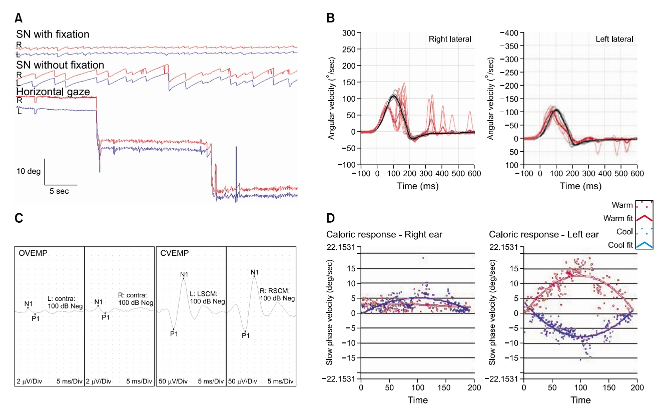
Fig. 1.(A) Video-oculography demonstrates spontaneous left beating nystagmus, suppressed by fixation. The horizontal gaze satisfies Alexander’s law, which has more degrees in the nystagmus direction. (red=right eye; blue=left eye). (B) Video head-impulse test revealed decreased gain (0.48) with covert and overt catch-up saccades in the right lateral semicircular canal. (C) Ocular vestibular evoked myogenic potential (VEMP) induced by sound stimulation (500-Hz tone burst) show delayed latency in the right ear of the patient, but indicated normal response in the left ear. Cervical VEMP show normal responses at both sides. (D) Caloric test revealed caloric paresis on the right side with 72%. SN, spontaneous nystagmus.


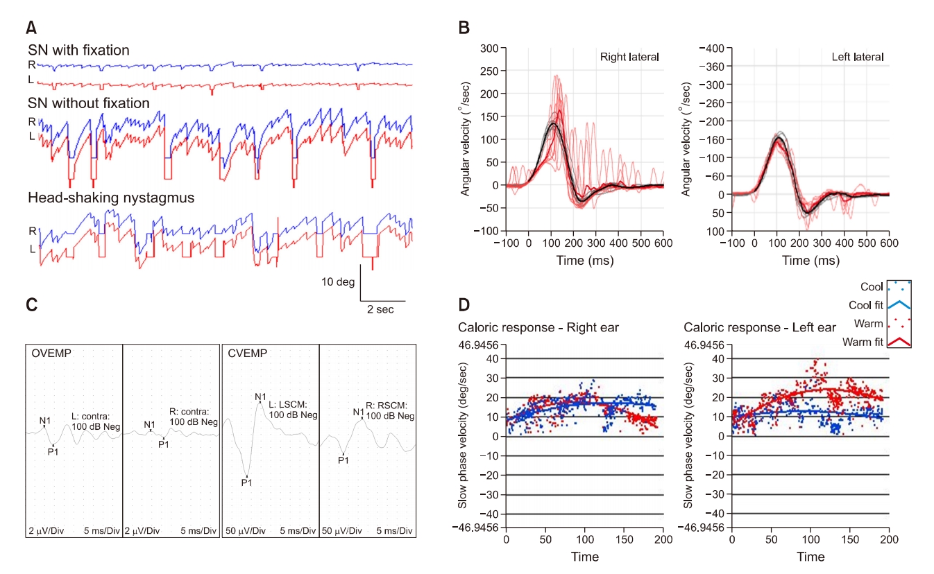
Fig. 2.(A) Video-oculography demonstrates spontaneous left beating nystagmus, suppressed by fixation. After head-shaking, the degree of nystagmus was more severe and did not change direction. (red=right eye; blue=left eye). (B) Video head-impulse test reveals decreased gain (0.44) with covert and overt catch-up saccades in the left lateral semicircular canal. (C) Ocular vestibular evoked myogenic potential (VEMP) test indicated normal response in the left ear but delayed latency in the right ear of the patient. Cervical VEMP show normal response at both sides. (D) Caloric test reveals caloric paresis on the right side with 79%. SN, spontaneous nystagmus.


- 1. Jeong SH, Kim HJ, Kim JS. Vestibular neuritis. Semin Neurol 2013;33:185–94.ArticlePubMedPDF
- 2. Halmagyi GM, Weber KP, Curthoys IS. Vestibular function after acute vestibular neuritis. Restor Neurol Neurosci 2010;28:37–46.ArticlePubMed
- 3. Bigelow RT, Agrawal Y. Vestibular involvement in cognition: visuospatial ability, attention, executive function, and memory. J Vestib Res 2015;25:73–89.ArticlePubMed
- 4. Kessels RP, Jaap Kappelle L, de Haan EH, Postma A. Lateralization of spatial-memory processes: evidence on spatial span, maze learning, and memory for object locations. Neuropsychologia 2002;40:1465–73.ArticlePubMed
- 5. Groth-Marnat G, Teal M. Block design as a measure of everyday spatial ability: a study of ecological validity. Percept Mot Skills 2000;90:522–6.ArticlePubMed
- 6. Lichtenberger EO, Kaufman AS. Essentials of WAIS-IV assessment. Hoboken (NJ): John Wiley & Sons; 2009.
- 7. Rönnlund M, Nilsson LG. Adult life-span patterns in WAIS-R Block Design performance: cross-sectional versus longitudinal age gradients and relations to demographic factors. Intelligence 2006;34:63–78.Article
- 8. van Asselen M, Kessels RP, Neggers SF, Kappelle LJ, Frijns CJ, Postma A. Brain areas involved in spatial working memory. Neuropsychologia 2006;44:1185–94.ArticlePubMed
- 9. Becker-Bense S, Dieterich M, Buchholz HG, Bartenstein P, Schreckenberger M, Brandt T. The differential effects of acute right- vs. left-sided vestibular failure on brain metabolism. Brain Struct Funct 2014;219:1355–67.ArticlePubMed
REFERENCES
Figure & Data
References
Citations
Citations to this article as recorded by 

- The Differential Effects of Acute Right- vs. Left-Sided Vestibular Deafferentation on Spatial Cognition in Unilateral Labyrinthectomized Mice
Thanh Tin Nguyen, Gi-Sung Nam, Jin-Ju Kang, Gyu Cheol Han, Ji-Soo Kim, Marianne Dieterich, Sun-Young Oh
Frontiers in Neurology.2021;[Epub] CrossRef
- Figure
- We recommend
- Related articles
-
- Dizziness in Patients with Vestibular Epilepsy
- Impairment of Vestibular Function in Patients with Vestibular Schwannoma According to the Presence of Dizziness
- Visuospatial dysfunction in patients with the right vestibular neuronitis
- The Clinical Efficacy of Vestibular Function Tests in Patients with Acute Unilateral Vestibulopathy
- The clinical characteristics of dizzy patients with normal vestibular function tests

 KBS
KBS
 PubReader
 ePub Link
 Cite
Cite


