척수소뇌실조 28형 1예
A Case of Spinocerebellar Ataxia Type 28
Article information
Abstract
Abstracts
Spinocerebellar ataxia type 28 (SCA 28) is characterized by young-adult onset, very slowly progressive gait and limb ataxia, dysarthria, nystagmus, ptosis, and ophthalmoplegia. It is caused by a heterozygous pathogenic mutation in the AFG3L2. So far, approximately 80 cases with genetically-confirmed SCA 28 have been reported in the literature. We report a patient with mild gait ataxia and dysarthria who carried a known pathogenic mutation in the AFG3L2. This is the first report of genetically-confirmed SCA 28 in Korea.
서론
척수소뇌실조(spinocerebellar ataxia, SCA)는 보행실조, 안구운동장애, 구음장애 등과 같이 소뇌 및 척수, 뇌간의 기능 장애를 특징으로 하는 신경퇴행성 질환이다[1]. 보통 상염색체 우성 유전을 따르며, 원인 유전자에 따라 현재 48형까지 보고되고 있지만 CAG 삼핵산 반복서열로 발생하는 SCA 2, 3, 6형이 흔한 것으로 알려져 있다. 저자들은 가족력이 뚜렷하지 않으면서 가벼운 보행실조 증상만보인 SCA 28형을 진단하였기에 문헌 고찰과 함께 보고하고자 한다.
증례
47세 여자가 1년 전부터 발생한 구음장애로 병원에 왔다. 처음에는 주로 피곤할 때 말이 어눌한 증상이 수 시간 정도 지속되다 호전되었지만, 내원 2개월 전부터는 호전없이 지속되었고, 계단이나 경사진 곳을 내려갈 때 중심을 잡기 힘든 증상이 발생하였다. 과거력에서 특이 병력은 없었고, 가족력에서도 소뇌실조증의 병력은 없었다. 신경학적 진찰에서 구음장애와 주시유발안진이 관찰되었지만 안구운동 범위, 신속안구운동, 원활추종안구운동검사는 정상이었다. 근력 및 감각기능 역시 정상이었고, 떨림(tremor)이나 경축(rigidity)과 같은 추체외로(extrapy-ramidal) 증상도 관찰되지 않았다. 소뇌기능검사에서 손가락-코검사(finger-to-nose test)와 발꿈치-정강이검사(heel- to-shin test) 시 측정이상(dysmetria)은 없었지만, 일자보행(tandem gait)시 양쪽으로 흔들리는 보행실조가 관찰되었다. 심부건반사는 하지에서 저하되어 있었고, 바빈스키 징후는 음성이었다.
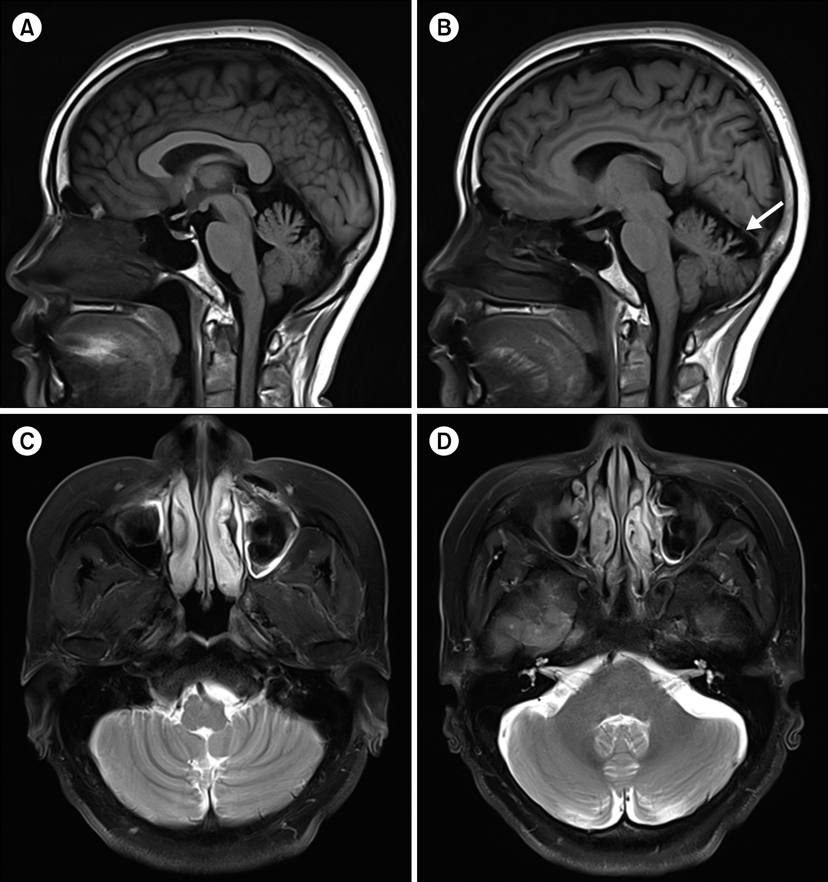
일반혈액검사, 화학검사, 갑상선기능 및 항체, 비타민 B12, 자가면역항체와 신생물딸림증후군(paraneoplastic synd-rome) 항체검사는 모두 정상이었다. 비디오안구운동검사에서 수평 주시유발안진은 관찰되었지만 자발안진이나 체위안진은 없었다. 신속안구운동과 원활추종안구운동은 정상이었다. 비디오 두부충동검사와 온도안진검사, 회전의자검사에서 전정안반사 기능은 정상이었다. 안저 촬영 및 안구 광학단층촬영에서 시신경 위축은 없었다. 뇌 자기공명영상(magnetic resonance imaging, MRI)에서 소뇌의 등쪽벌레(dorsal vermis)에 경미한 위축이 관찰되었지만 소뇌반구(cerebellar hemisphere)와 뇌간은 상대적으로 잘 보존되어 있었다(Fig. 1). 소뇌실조증의 가족력은 없었지만 진찰에서 구음장애, 주시유발안진, 경도의 보행실조 등 소뇌기능장애가 관찰되었고, 검사에서는 기질적 질환을 의심할 만한 소견이 확인되지 않아 SCA를 감별하기 위해 유전자 검사를 시행하였다. 우선 SCA 1, 2, 3, 6, 7, 8, 17형을 감별하기 위해 CAG 삼핵산 반복서열을 확인했지만 모두 정상 범위였다. 이후 소뇌실조 유전자 패널을이용한 차세대염기서열분석을 시행한 결과 AFG3L2 유전자에서 이형접합 과오돌연변이인 c.1977T> C (p.Met666Thr, NM_006796.2)를 확인할 수 있었다(Fig. 2). 이는 국외에서 SCA 28형의 병인으로 보고된 유전자 변이(rs151344515)로, 한국인 참조 유전체 데이터베이스(Korean Reference Genome Database)에서 정상인에서는 존재하지 않았다[2].

Brain magnetic resonance imaging of the patient. Sagittal T1-weighted images (A, B) exhibit mild atrophy of the cerebellar ver-mis (white arrow), but axial T2- weighted images (C, D) show a rel-ative sparing of the cerebellar hemi-sphere.

Sequencing result of the missense mutation in the AFG3L2 identified by targeted next-generation sequencing. The chromato-gram shows a heterozygous missense mutation in exon 16 causing the substitution of the highly conserved methionine by threonine at position 666 (c.1997T>C [p.Met666Thr]). This has been regis-tered in the Single Nucleotide Polymorphism Database (dbSNP) as a known pathogenic mutation (rs151344515).
현재까지 4년 동안 추적 관찰 중이지만 구음장애와 보행실조 증상은 큰 변화 없이 지속되고 있으며, 사지실조나 안구운동장애 등은 관찰되지 않고 있다.
고찰
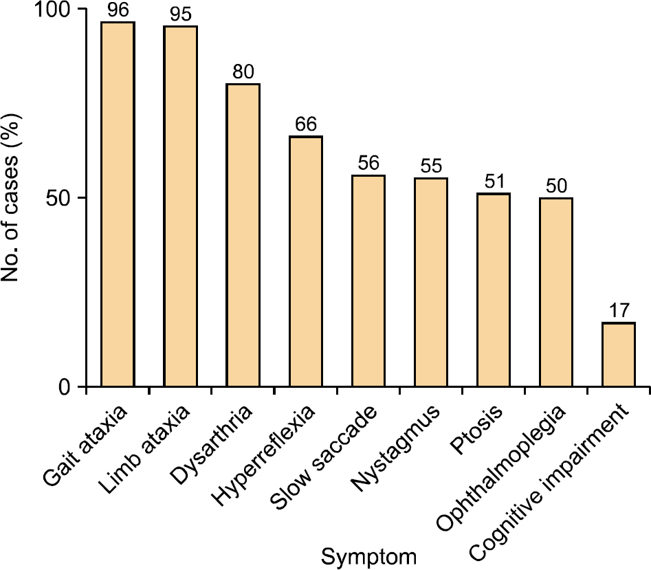
SCA 28형은 염색체 18p11에 위치한 AFG3L2 유전자 변이로 발생하는 질환이다[3]. 상염색체 우성 소뇌실조(autosomal dominant cerebellar ataxia)의 1.5% 정도로 매우 드물어 국외에서는 현재까지 80예 정도 보고되고 있지만 국내에서는 아직 보고된 적이 없다[2]. 현재까지 보고된 증례들을 살펴보면, 평균 발병 나이는 31세이지만 3세에서 70세까지 다양한 연령대에서 발병할 수 있다[2–7]. 대부분의 환자에서 보행실조(96%)와 사지실조(95%), 구음장애(80%)가 발생하지만, 평균 20년 정도의 이환 기간 동안 대부분(90%) 독립보행이 가능할 정도로 병의 진행이 느렸다. 실조 증상 외 심부건 반사항진(66%), 느린 신속보기(56%), 주시유발안진(55%), 눈꺼풀처짐(51%), 안근마비(50%), 인지기능 장애(17%) 등을 보일 수 있다(Fig. 3). MRI 에서 소뇌 위축은 약 77%로 보고되지만, 주로 벌레(ver-mis) 부위의 위축이 관찰되었다. 본 증례에서는 보행실조와 구음장애, 주시유발안진 외 다른 증상은 보이지 않았지만 이환 기간이 4년밖에 되지 않았기 때문에 향후 다른증상들이 발생할 가능성이 있다.
AFG3L2 유전자는 사립체 내막(inner mitochondrial mem-brane)에 위치하는 m-AAA 사립체 단백질 분해효소(mito-chondrial protease)를 부호화(encoding)한다[4]. 이 단백질 분해효소는 사립체에서 단백질의 합성과 분해를 조절하여 단백질의 항상성 유지에 중요한 역할을 한다. 구조적으로는 AFG3L2 단백질로만 구성된 homo-oligomeric 복합체와, SPG7 유전자의 산물인 paraplegin 단백질과 함께 구성된 hetero-oligomeric 복합체로 나눌 수 있다. AFG3L2와 para-plegin 단백질은 소뇌의 푸르키네세포(Purkinje cell)와 심부 소뇌핵(deep cerebellar nuclei)에 위치하고 있어 이들 유전자에 변이가 발생하면 푸르키네세포 내 단백질 합성 장애로 소뇌실조가 발생한다. 특히 paraplegin 단백질은 척수 운동신경 세포에도 많이 분포되어 있어, SPG7 유전자 변이는 강직하반신마비 7형(spastic paraplegia type 7)과 연관되어 있다[4]. 또한 AFG3L2 유전자는 변이의 위치에 따라 임상형이 다른 것으로 알려져 있다. AFG3L2 단백질 내 중요한 도메인은 AAA 도메인(아미노산 344–477)과 peptidase M41 도메인(아미노산 541–744)이다. 이 중 SCA 28형은 peptidase M41 도메인의 변이와 연관되어 있는 반면, AAA 도메인의 변이는 상염색체 우성 시신경위축(dominant optic atrophy)을 일으킨다[8,9]. 본 증례의 경우 peptidase M41 도메인에 AFG3L2 유전자 변이가 위치하고 있어 시신경위축 없이 소뇌실조 증상만 관찰되었다.
SCA28은 아주 느리게 진행하는 실조증상이 주요한 특징이지만, 안근마비와 눈꺼풀처짐 역시 비교적 흔하게 관찰된다. 문헌 고찰에서도 약 50%의 환자에서 관찰되었으며, 만성 진행외안근마비(chronic progressive external oph-thalmoplegia)로 오인된 경우도 있었다[2–7]. 대부분 이환 기간이 평균 25년 정도 경과한 후 안근마비와 눈꺼풀처짐이 확인되었기 때문에 병이 진행함에 따라 안근마비나 눈꺼풀처짐이 발생함을 시사한다. 본 증례의 경우 이환 기간이 4년밖에 되지 않았기 때문에 안근마비와 눈꺼풀처짐이 관찰되지 않았을 가능성이 크다. SCA에서 안근마비는 흔히 관찰되는 신경학적 징후이다. 특히 SCA 2형과 7형에서는 초기에 느린 신속보기가 발생하고, 이후 병이 진행함에 따라 안근마비가 발생한다. 대부분 안구운동에 관여하는 뇌간 구조물의 장애로 인한 핵상마비(supra-nuclear palsy)가 주 원인으로 여겨지지만, SCA 28형에서는 외안근의 위축도 안근마비에 기여하는 것으로 알려져 있다[10]. 이는 SCA 28형이 사립체 기능 이상과 연관되어 있으며, 외안근이 사립체 질환에 취약하기 때문이다.
SCA 28형은 실조증상이 심하지 않고 아주 느리게 진행하기 때문에 임상적으로 간과할 수 있다. 하지만 최근 유전자 진단법의 발달로 소뇌실조증의 유전형 진단이 비교적 수월해지고 있다. 따라서 소뇌실조의 이환 기간이 비교적 길지만, 독립보행이 가능하고 안근마비나 눈꺼풀처짐이 동반되어 있을 경우에는 SCA 28형의 가능성을 염두에 두어야 한다.
중심 단어: 척수소뇌실조 28형, AFG3L2 유전자, 보행실조
이해관계(CONFLICT OF INTEREST)
저자들은 이 논문과 관련하여 이해관계의 충돌이 없음을 명시합니다.
감사의글(ACKNOWLEDGMENTS)
본 연구는 2020년도 양산부산대학교병원 임상연구비 지원으로 이루어졌습니다.

