Skip Navigation
Skip to contents

Res Vestib Sci : Research in Vestibular Science

OPEN ACCESS
SEARCH
Search

Articles

Page Path
HOME > Res Vestib Sci > Volume 17(4); 2018 > Article
Case Report
경막외 신경차단술 시행 중 발생한 감각신경성난청과 어지럼 1예
이병민1, 노진홍2, 안성기1, 박현우1orcid
A Case of Sensorineural Hearing Loss and Vertigo during Epidural Nerve Block
Byeong Min Lee1, Jin hong Noh2, Seong Ki Ahn1, Hyun Woo Park1orcid
Research in Vestibular Science 2018;17(4):170-174.
DOI: https://doi.org/10.21790/rvs.2018.17.4.170
Published online: December 21, 2018

1Department of Otorhinolaryngology-Head and Neck Surgery, Gyeongsang National University Hospital, Jinju, Korea

2Department of Neurology, New Tongyoung Hospital, Tongyoung, Korea

Corresponding Author: Hyun Woo Park Department of Otorhinolaryngology-Head and Neck Surgery, Gyeongsang National University Hospital, 79 Gangnam-ro, Jinju 52727, Korea Tel: +82-55-750-8178 Fax: +82-55-759-0613 E-mail: phw83@hanmail.net
• Received: October 11, 2018   • Revised: December 3, 2018   • Accepted: December 4, 2018

Copyright © 2018 by The Korean Balance Society. All rights reserved.

This is an open access article distributed under the terms of the Creative Commons Attribution Non-Commercial License (http://creativecommons.org/licenses/by-nc/4.0) which permits unrestricted non-commercial use, distribution, and reproduction in any medium, provided the original work is properly cited.

  • 4,988 Views
  • 78 Download
prev
  • Epidural anesthesia has significantly advanced in neuraxial anesthesia and analgesia. It is used for surgical anaesthesia and treatment of chronic pain. Hearing loss during or after epidural anesthesia is rare, and it is known to occur by the change of the intracranial pressure. Cerebrospinal fluid is connected with perilymph in the cochlear and vestibule that is important to hearing and balance. If the intracranial pressure is abruptly transferred to the inner ear, perilymph can be leak, that called perilymphatic fistula, dizziness, and hearing loss can occur suddenly. We report a 65-year-old woman who presented with acute onset dizziness and hearing loss during the epidural nerve block for back pain, wherein we speculated a possibility of perilymphatic fistula as the mechanism of hearing loss and dizziness. The mechanism of dizziness and hearing loss was suspected with perilymphatic fistula.
경막외 마취(epidural anesthesia)는 가장 흔한 국소 마취기법 중 하나로 알려져 있으며 주로 정형외과, 산부인과, 비뇨기과, 일반외과 등에서 시행하는 수술에 많이 사용되고있다. 또한 경막외 신경차단술(epidural nerve block)은 추간판탈출증(intervertebral disk herniation), 척추관절강직증(spondylosis), 신경근병(radiculopathy), 척추관협착증(spinal stenosis) 등으로 인한 만성 통증의 조절을 위해서도 이용되고 있다.
이러한 다양한 사용에도 불구하고 두통이나 저혈압, 심정지, 하지마비, 발작, 신경학적 손상, 신경근병, 말총증후군(cauda equine syndrome) 그리고 사망에 이르기까지 다양한 합병증이 나타날 수 있다. 그러나 이런 합병증의 발생률은 상당히 낮으며 0.1% 미만으로 보고되고 있다[1]. 특히 청력 소실이나 어지럼과 같은 청각전정계(audiovestibular system) 이상은 0.2%–0.8%에서 보고되고 있으나 증상은 대게 일시적으로 나타난다고 알려져 있다[2].
경막외 차단술로 인한 청각전정계 이상은 수두증(hydrocephalus), 미세색전(microemboli), 감염으로 그 기전이 설명될 수 있으며 뇌척수액의 소실로 인해 외림프의 압력이 낮아지면서 발생하는 내림프수종(endolymphatic hydrops)으로도 나타날 수 있다고 알려져 있다[3-5].
요추 천자 시 천자침이 극돌간인대(interspinous ligament)를 지나 삽입될 때 생리 식염수나 공기로 채워진 주사기를 연결하게 된다. 천자침을 계속 진입시키면 황색인대(ligamentum flavum)에 닿게 되는데 이는 주사기 내용물 주입 시 저항이 느껴지는 것을 통해 알 수 있다. 천자침이 경막외 공간으로 진입하게 되면 생리 식염수나 공기가 저항 없이 쉽게 주입되게 되는데 이를 통해 경막외 공간을 확인할 수 있으며 이런 방법을 마취통증의학에서 ‘loss of resistance technique’ 이라 한다[6]. 이런 기법을 이용하다 의도했던 것보다 많은 양의 생리 식염수나 공기가 경막외 공간으로 주입되게 되면 두개 내 압력이 올라가게 되고 높아진 압력은 와우도수관(cochlear aqueduct)을 통해 외림프공간으로 전달될 수 있으며 이런 일련의 과정은 청력 소실이나 어지럼을 일으킬 수 있는 외림프누공(perilymphatic fistula)을 발생 시킬 수 있을 것이다[7].
저자들은 경막외 신경차단술 시행 중 loss of resistance technique중에 과주입된 공기에 의해서 발생한 감각신경성 난청과 어지럼으로 생각되는 증례를 경험하였기에 문헌고찰과 함께 보고하는 바이다.
65세 여자 환자가 척추관협착증으로 인한 만성 통증을 조절하기 위해 타병원 신경외과에 내원하여 경막외 신경차단술을 받았다.
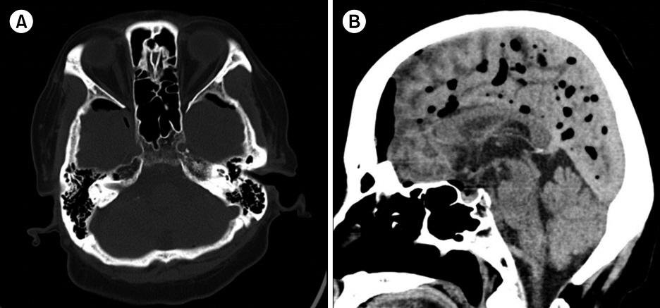
시술을 진행하면서 술자는 경막외 공간을 확인하기 위해 공기를 주입하였는데 이때 환자는 왼쪽 귀에서 폭발성의 소리와 함께 두통, 심한 회전성어지럼, 좌측 청력 소실과 이명이 발생하였다고 하였으며 이와 같은 증상 발생 후 즉시 시술은 중단되었고 절대안정을 취하며 본원 이비인후과로 의뢰되었다. 증상이 발생한 직후 시행한 뇌 computed tomography (CT)상 많은 양의 공기뇌증(pneumocephalus)이 관찰되었으며 뇌 실질 내의 다른 특이 소견은 보이지 않았다(Fig. 1).
이경 검사상 양쪽 고막은 정상이었으며 Frenzel 고글검사상 자발안진이 우측으로 관찰되었고 두진(head shaking) 이후 더욱 커지는 양상을 보였다. 두위안진(positional nystagmus) 및 소뇌기능검사(cerebellar function test)상 이상 소견은 보이지 않았으며 순음청력검사상 우측 역치는 정상범위였으나 좌측은 농 상태였다(Fig. 2). 전정기능검사 결과 양온교대안진검사(bithermal caloric test)상 좌측에서 33% 반고리관 마비(canal paresis)가 확인되었고 누공검사(fistula test)를 포함하여 다른 항목에서는 이상 소견을 보이지 않았다(Fig. 3). 저자들은 이학적 검사에서는 특이 소견이 보이지 않으나 과거력을 고려했을 때 경막외 신경차단술 시 주입된 공기로 인한 외림프누공이 의심되었다. 환자 및 보호자에게 상태를 설명하고 시험적 고실개방술(exploratory tympanotomy)을 권유하였으나 수술적 치료는 거부하여 절대안정, 약물치료 및 보존적 치료를 시행하였다. 약물치료는 고용량 경구 스테로이드 치료(methylprednisolone 0.8 mg/kg)를 10일간 시행하였고 고실 내 스테로이드 주입술(dexamethasone 5 mg)을 2주간 4회 시행하였으며 전정 재활 치료도 약물치료와 함께 진행하였다. 증상 발생 2주 뒤에 시행한 뇌 CT상 공기뇌증은 호전되었으며 다른 뇌실질에 이상 소견은 관찰되지 않았다.
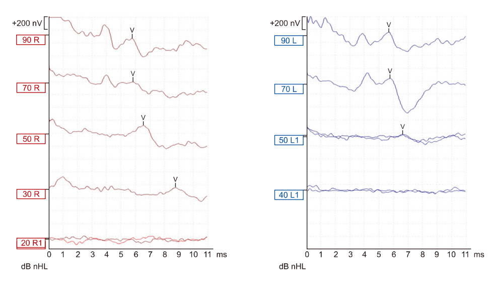
이후 적극적인 전정 재활치료로 환자의 어지럼은 호전되었으며 자발안진 및 두진 후 안진 또한 관찰되지 않았고 발생 1개월 후 시행한 청성뇌간반응검사(auditory brainstem response)상 좌측 청력은 50 dB로 회복되었다(Fig. 4).
요추천자나 경막외 마취와 관련된 청력 소실은 1914년 처음 보고가 되었으며 두개 내 압력 상승으로 인한 정원창누공은 1970년에 Stroud와 Calcaterra가 처음으로 보고하였다. 이후 여러 문헌들을 통해 두개 내 압력이 상승될 경우 지주막하 공간과 와우도수관을 통해 높아진 압력이 내이로 전달되게 되고 이로 인해 외림프누공이 발생할 수 있다고 알려졌다[8].
외림프누공은 림프액으로 채워진 내이와 공기로 채워진 중이 사이에 교통이 생긴 상태를 말하며 외림프누공이 발생했을 때에는 초기에 적절한 치료가 이루어지지 않으면 영구적인 난청과 조절되지 않는 어지럼이 남을 수 있다.
병태생리로는 외측폭발이론(explosive theory)과 내측폭발이론(implosive theory)으로 설명하고 있다. 외측폭발이론의 경우 이번 증례처럼 상승된 두개 내 압력이 와우도수관을 통해 내이로 전달되게 되면 이로 인해 난원창 막의 파열이나 등골 윤상 인대의 파열로 나타날 수 있다는 것이고 내측폭발이론은 발살바 조작이나 대기압의 급격한 변화로 인해 중이강 내의 압력이 상승되게 되면 이는 정원창 막을 내측으로 파열시키거나 등골 족판이 내측으로 탈구된다는 기전으로 설명하고 있다[9]. 그리고 외측폭발이나 내측폭발 이외에 종양이나 외상 또는 이과적 수술로 인한 직접적인 손상으로도 발생할 수 있다[10].
두개내압 상승으로 인한 외림프누공 이외에 청력 소실과 어지럼을 설명할 수 있는 다른 가설로는 내이신경(vestibulocochlear nerve)의 늘어짐, 내이도 내부로 팽장한 뇌수막류(meningocele), 뇌척수액 손실로 인한 내림프수종 가능성 등도 보고되어 있으나 이번 증례에서는 환자가 느낀 폭발성 소리와 직후 발생한 어지럼과 난청을 고려했을 때 외림프누공이 가장 의심되었다[11,12].
비록 이번 증례에서 누공검사를 포함한 이학적검사에서 특이 소견이 없었으며 영상의학적 검사상에서도 기전정(pneumolabyrinth)이나 중이강 내의 액체 저류를 확인하지는 못하였으나 발생 당시 환자의 증상을 바탕으로 생각해 보았을 때 경막외 공간의 확인을 위해 시행한 ‘loss of resistance technique’ 중에 주입된 공기가 두개 내 공간으로 들어가 두개 내 압력을 증가시켰고, 이 압력이 내이로 전달되어 외림프누공을 발생시켜 급작스런 두통이나 폭발적인 소리, 청력 소실과 어지럼이 생겼을 것이라 생각하였다.
외림프누공의 치료 목적은 청력의 회복과 어지럼의 호전이다. 첫 번째는 이번 증례처럼 침대 머리 쪽을 30도 정도 거상하여 5일에서 7일간 침상안정을 취하며 경과를 보는 것이다. 그 동안 발살바 조작을 하지 않도록 교육해야 하고 안정제, 항현훈제, 하제 등을 투여하여 증상 완화에 도움을 줄 수 있으며 이런 비 수술적 방법으로 30%–40%에서 호전될 수 있다. 만약 비수술적 방법으로 호전이 없거나 증상이 더욱 악화되는 경우에는 수술적 치료를 고려해야 한다. 수술은 진단과 치료 목적으로 시험적 고실개방술을 시행해 볼 수 있으며 정원창 난원창 주변에 액체 저류가 있거나 외림프누공을 직접 확인함으로써 외림프누공을 확진함과 동시에 치료할 수 있다[13].
경막외 마취 또는 경막외 신경차단술은 경막외 공간에 약물을 주입함으로써 통증의 전달을 막게 되고 이를 통해 통증 조절이나 수술적 치료에 필요한 마취를 목적으로 사용될 수 있다. 이 술기를 시행할 때 경막외 공간의 확인을 위해 이용하는 ‘loss of resistance technique’ 기법은 시술 시에 술자로 하여금 과도한 생리 식염수나 공기가 주입되지 않도록 주의가 필요하며 만약 술기 직후 환자가 청력 소실이나 어지럼을 호소한다면 외림프누공을 의심하고 적절한 이학적 검사 및 영상의학적 검사를 통해 감별할 수 있어야 하며 일단 의심이 될 경우에는 적극적인 치료를 통해 영구적인 청력 소실을 막아야 한다. 이번 증례에서는 두개내압 증가에 의한 외측폭발 기전으로 발생한 외림프누공이 강력히 의심되었으나 환자의 스트레스와 불안으로 시험적 고실개방술을 시행하지 못한 것이 가장 큰 한계라고 생각한다. 만약 이와 같거나 비슷한 경우가 발생하면 적극적이고 빠른 대처가 필요할 것이다.

저자들은 이 논문과 관련하여 이해관계의 충돌이 없음을 명시합니다.

Fig. 1.
Brain computed tomographic image. Diffuse pneumocephalus. Axial view (A) and sagittal view (B).
rvs-17-4-170f1.jpg
Fig. 2.
Pure tone audiogram after hearing loss. Hearing threshold was 20 dB at right side (A) and no response at left side (B).
rvs-17-4-170f2.jpg
Fig. 3.
Caloric test in vestibular function test. Left caloric response was 33% weaker than right. SCV, slow component velocity; avg, average; FI, fixation index; F, Fixation; NF, nonfixation.
rvs-17-4-170f3.jpg
Fig. 4.
Auditory brainstem response after treatment. Hearing threshold was 50 dB at left side. nHL, normalized hearing level.
rvs-17-4-170f4.jpg
  • 1. Auroy Y, Narchi P, Messiah A, Litt L, Rouvier B, Samii K. Serious complications related to regional anesthesia: results of a prospective survey in France. Anesthesiology 1997;87:479–86.ArticlePubMed
  • 2. Kiliçkan L, Gürkan Y, Ozkarakas H. Permanent sensorineural hearing loss following spinal anesthesia. Acta Anaesthesiol Scand 2002;46:1155–7.ArticlePubMed
  • 3. Sundberg A, Wang LP, Fog J. Influence of hearing of 22 G Whitacre and 22 G Quincke needles. Anaesthesia 1992;47:981–3.ArticlePubMed
  • 4. Stoeckli SJ, Böhmer A. Persistent bilateral hearing loss after shunt placement for hydrocephalus. Case report. J Neurosurg 1999;90:773–5.ArticlePubMed
  • 5. Wang LP, Fog J, Bove M. Transient hearing loss following spinal anaesthesia. Anaesthesia 1987;42:1258–63.ArticlePubMed
  • 6. Saberski LR, Kondamuri S, Osinubi OY. Identification of the epidural space: is loss of resistance to air a safe technique? A review of the complications related to the use of air. Reg Anesth 1997;22:3–15.ArticlePubMed
  • 7. Ciuman RR. Communication routes between intracranial spaces and inner ear: function, pathophysiologic importance and relations with inner ear diseases. Am J Otolaryngol 2009;30:193–202.ArticlePubMed
  • 8. Conde M, Villanueva A, Sánchez P, Pérez JA, Ruiz-Rico R. Deafness secondary to lumbar puncture. Rev Neurol 1997;25:1218–20.PubMed
  • 9. Goodhill V. Sudden deafness and round window rupture. Laryngoscope 1971;81:1462–74.ArticlePubMed
  • 10. Al Felasi M, Pierre G, Mondain M, Uziel A, Venail F. Perilymphatic fistula of the round window. Eur Ann Otorhinolaryngol Head Neck Dis 2011;128:139–41.ArticlePubMed
  • 11. Cosar A, Yetiser S, Sizlan A, Yanarates O, Yildirim A. Hearing impairment associated with spinal anesthesia. Acta Otolaryngol 2004;124:1159–64.ArticlePubMed
  • 12. Truesdale CM, Peterson RB, Hudgins PA, Vivas EX. Internal auditory canal meningocele-perilabyrinthine/translabyrinthine fistula: case report and imaging. Laryngoscope 2016;126:1931–4.ArticlePubMed
  • 13. Kohut RI, Hinojosa R, Ryu JH. Update on idiopathic perilymphatic fistulas. Otolaryngol Clin North Am 1996;29:343–52.ArticlePubMed

Figure & Data

References

    Citations

    Citations to this article as recorded by  

      • PubReader PubReader
      • ePub LinkePub Link
      • Cite
        CITE
        export Copy
        Close
        Download Citation
        Download a citation file in RIS format that can be imported by all major citation management software, including EndNote, ProCite, RefWorks, and Reference Manager.

        Format:
        • RIS — For EndNote, ProCite, RefWorks, and most other reference management software
        • BibTeX — For JabRef, BibDesk, and other BibTeX-specific software
        Include:
        • Citation for the content below
        A Case of Sensorineural Hearing Loss and Vertigo during Epidural Nerve Block
        Res Vestib Sci. 2018;17(4):170-174.   Published online December 21, 2018
        Close
      • XML DownloadXML Download
      Figure
      • 0
      • 1
      • 2
      • 3
      Related articles
      A Case of Sensorineural Hearing Loss and Vertigo during Epidural Nerve Block
      Image Image Image Image
      Fig. 1. Brain computed tomographic image. Diffuse pneumocephalus. Axial view (A) and sagittal view (B).
      Fig. 2. Pure tone audiogram after hearing loss. Hearing threshold was 20 dB at right side (A) and no response at left side (B).
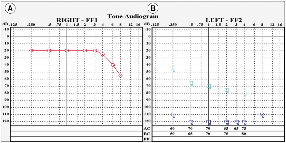
      Fig. 3. Caloric test in vestibular function test. Left caloric response was 33% weaker than right. SCV, slow component velocity; avg, average; FI, fixation index; F, Fixation; NF, nonfixation.
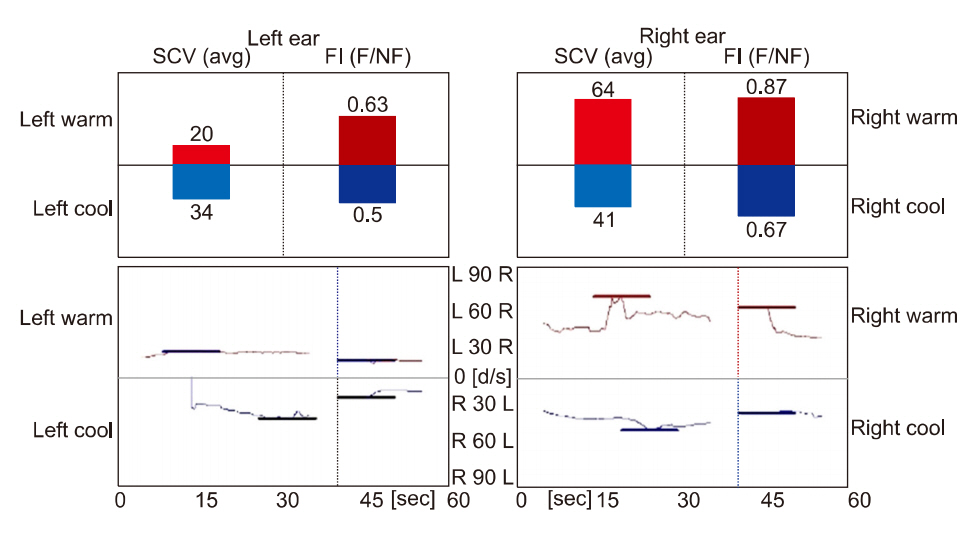
      Fig. 4. Auditory brainstem response after treatment. Hearing threshold was 50 dB at left side. nHL, normalized hearing level.
      A Case of Sensorineural Hearing Loss and Vertigo during Epidural Nerve Block

      Res Vestib Sci : Research in Vestibular Science
      TOP